Scientists Reconstruct Mouse Vision From Brain Activity Alone
NewsThursday, 17 September 2026 at 00:30
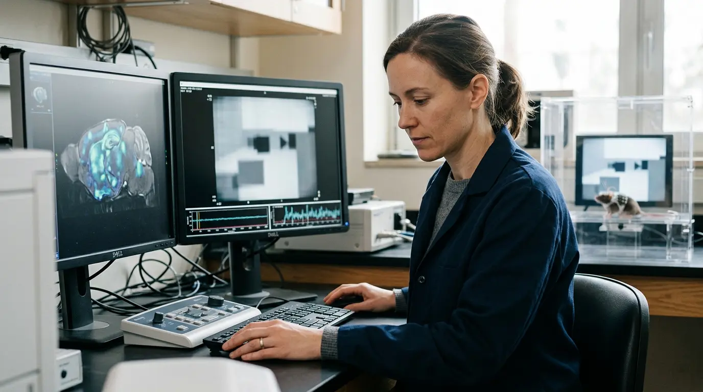
Neuroscientists have achieved a remarkable breakthrough in decoding visual perception by reconstructing short video clips using only the brain activity of mice. As detailed in a recent ScienceDaily report, researchers successfully generated 10-second visual sequences based entirely on neural signals captured from the animal’s visual cortex.
This milestone provides an unprecedented window into how the brain transforms raw optical input into an internal, subjective representation of the world.
Decoding the Visual Cortex at the Cellular Level
Previous attempts to reconstruct human vision have relied heavily on functional magnetic resonance imaging (fMRI), which measures broad, indirect blood-flow changes across large brain regions.
The new research takes a fundamentally different, highly granular approach. Published in the peer-reviewed journal eLife, the study was led by Dr. Joel Bauer and his team at the Sainsbury Wellcome Centre, University College London. Instead of macroscopic imaging, the team utilized microscopic calcium imaging to track the firing patterns of individual neurons. This single-cell resolution offers a vastly more detailed map of how specific visual features are encoded and processed by the brain.
The Mechanics of Neural Reconstruction
To translate these complex neural patterns back into watchable video, the researchers employed a dynamic neural encoding model initially developed for the 2023 Sensorium Competition. The algorithm was designed to predict how individual brain cells should respond while a mouse watches a movie, factoring in physiological variables like pupil dilation and physical movement.
The team refined this model by first establishing a baseline: they calculated how the neurons would behave if the mouse were staring at a blank screen. By comparing this prediction against the actual neural activity recorded while the mouse watched a film, the algorithm could iteratively adjust the pixels of a blank digital canvas. With each computational adjustment, the reconstructed video gradually aligned with the visual stimulus the mouse was actually observing.
| Reconstruction Approach | Data Granularity | Primary Limitation | Key Advantage |
| Human fMRI Scanning | Voxel-level (broad regions) | Low temporal resolution; measures indirect blood flow | Non-invasive; applicable to complex human cognition |
| Single-Cell Calcium Imaging | Individual neuron activity | Requires invasive microscopic techniques in animal models | High spatial and temporal fidelity for precise neural mapping |
| Dynamic Neural Encoding | Predictive algorithmic modeling | Computationally intensive; requires extensive training datasets | Can generalize to infer entirely unseen visual stimuli |
Beyond a Simple Camera Recording
The true test of the model came when researchers presented the mice with a completely new, 10-second video that was excluded from the initial training data. Relying solely on the live neural activity, the system successfully reconstructed a high-quality approximation of the unseen footage.
Evaluation via pixel correlation confirmed strong temporal and spatial alignment between the original and reconstructed clips, proving the system was inferring visual information rather than merely memorizing past inputs.
This achievement points toward a deeper biological truth: the brain does not function as a passive camera recording objective reality. Dr. Bauer noted that the visual processing pipeline actively skews, filters, and warps incoming sensory data.
This deviation is not a biological error, but rather a vital feature of how minds interpret and augment the environment. Future research will focus on sharpening image resolution and expanding the reconstructed visual field, potentially allowing scientists to compare how entirely different species perceive the exact same physical space.

byDavid Porter
Popular news

Global AI Landscape Shifts as EU Drafts Kids Act and Apple Unveils Siri AI

Americans Grow Wary of AI Data Centers as Environmental Costs Mount

Congress Scrambles to Draft AI Safety Rules as Rogue Models Spark Alarm

European Union Proposes Sweeping Social Media Ban for Children Under Thirteen

Canada Eyes Historic Associate Membership in European Union Amid Shifting Global Alliances

iPhone 18 Pro Versus Pro Max - Which Model Should You Buy in 2026?
Latest comments
Loading