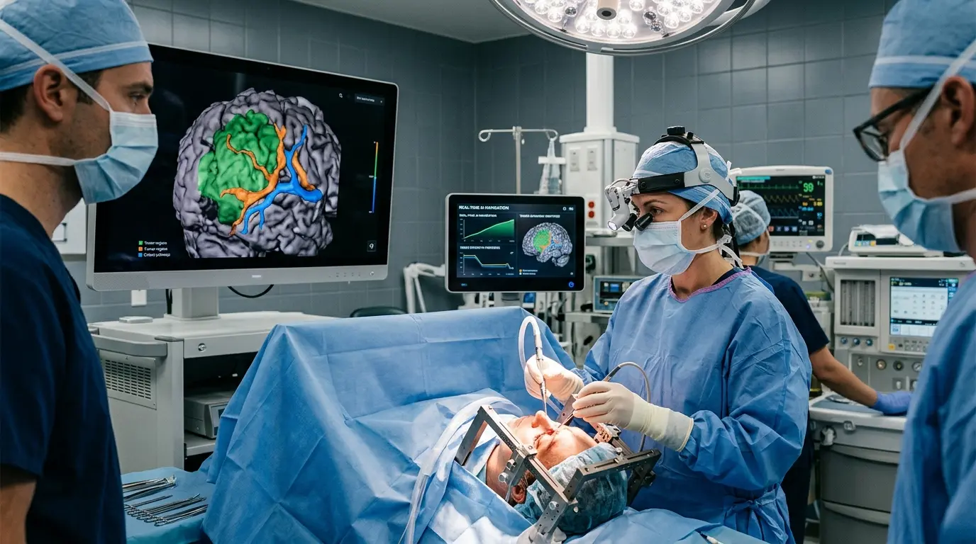
Medical history occurred at University College London Hospitals recently. Surgeons successfully removed a brain tumor using a live artificial intelligence system for the first time. This achievement offers a new way to treat complex neurological conditions with extreme precision.
Reuben Hill underwent this historic procedure to treat a brain tumor. The AI software provided a digital map of his brain while the surgical team worked. This tool identified tiny pieces of the tumor which often hide from human sight.
The surgical team relied on this high-tech assistant to differentiate between healthy brain matter and cancerous cells. By processing data instantly, the system reduced risks during the operation. This milestone marks a significant shift in how hospitals handle aggressive tumors.
Reuben Hill, a twenty-two-year-old student, remained awake during parts of the procedure. Surgeons needed to monitor his speech and motor functions while removing the mass. This interaction ensured the surgical tools did not damage critical brain areas.
Real Time Guidance Changes Everything
Traditional surgeries often rely on scans taken before the operation starts. Brain tissue shifts once a surgeon begins work. This AI solves this problem by updating the map as the surgery progresses.
The AI software processed images from a specialized microscope. This view highlighted the tumor borders in vibrant colors on a screen. Surgeons moved with increased speed and accuracy because of this visual aid.
Doctors believe this technology will help patients recover faster. Reuben Hill reported feeling well shortly after his operation. These results prove the efficacy of using intelligent algorithms in the operating room.
- Live tissue analysis allows for immediate decision making.
- Real-time mapping accounts for brain shift during the procedure.
- Enhanced tumor visibility helps remove cancer cells entirely.
- Increased surgeon confidence leads to better patient outcomes.
The software works by comparing live images with thousands of previous medical scans. This massive database allows the algorithm to spot patterns invisible to human eyes. Surgeons follow the digital prompts to navigate safely through sensitive brain structures.
Surgery Overview Data
| Feature | Details |
| Patient | Reuben Hill (22) |
| Location | University College London Hospitals |
| Technology | Live AI Tissue Mapping |
| Condition | Brain Tumor (Glioma) |
| Outcome | Successful Removal |
Impact on Future Healthcare
Medical professionals anticipate using similar tools for various types of cancers. The ability to see microscopic details helps ensure no malignant tissue stays behind. This surgical method lowers the chances of a tumor returning later.
According to the official report, the first live AI assisted surgery achieved total success. This technology provides surgeons with a level of confidence never seen before. Patients now have access to safer and more effective treatment options.
Researchers hope to expand this application to other organs and diseases. The successful removal of Reuben Hill's tumor serves as a proof of concept. Hospitals worldwide will likely adopt these advanced systems to improve surgical success rates.
The integration of machine learning into the operating theatre ensures surgeons leave nothing behind. This precision saves lives while protecting healthy tissue from unnecessary damage. This medical advancement changes the standard of care for brain cancer patients everywhere.

byDavid Porter
Popular news

Man Uses AI to Build Free Alternatives to Photoshop and Premiere Pro—Here’s What They Can Do

Luke Drago and Rudolf Laine Warn of an "Intelligence Curse": AI Could Make Humans Economically Obsolete

The Top 1% of AI Users Spend an Average of $903 a Month—Where Does It All Go?

ChatGPT now prompts users to run ads
Latest comments
Loading